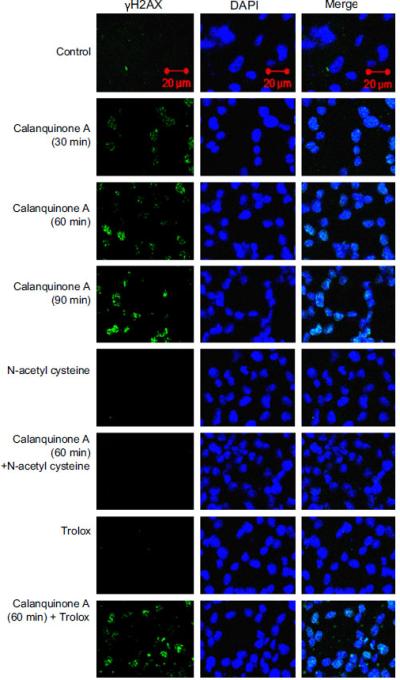
Fig. 3.

Effect of calanquinone A γH2A.X formation. A172 cells were incubated in the absence or presence of the indicated agent (calanquinone A, 4μM; N-acetyl cysteine, 1 mM; trolox 0.3 mM). The cells were fixed for the confocal fluorescence microscopic detection of γH2A.X formation (green fluorescence) in the nucleus (blue-fluorescent DAPI staining). (For interpretation of references to color in this figure legend, the reader is referred to the web version of this article.)
