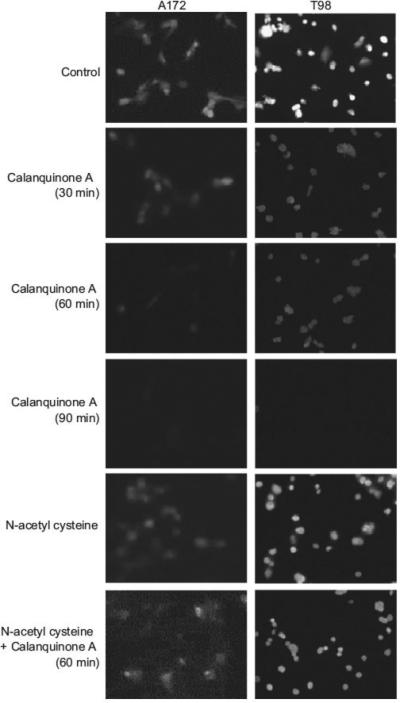
Fig. 5.

Effect of calanquinone A on cellular glutathione content. The cells were incubated in the absence or presence of the agent (calanquinone A, 4 μM; N-acetyl cysteine, 1 mM) for the indicated time. The cellular glutathione was detected by fluorescence microscopic examination of monochlorobimane staining. Data are representative of three independent experiments.
