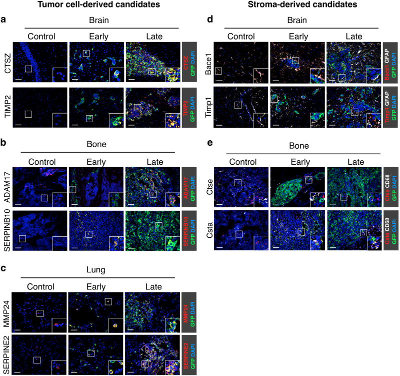
Figure 2. Independent validation of differentially expressed genes in experimental brain, bone and lung metastases.
(a-e) Representative images of control (non tumor-burdened) tissue, early- and late-stage site-specific metastases (classified by BLI intensity as in Supplementary Fig. 1; n=3 samples for each stage and tissue) showing immunofluorescence staining of tumor- and stromal-derived proteases and protease inhibitors exhibiting stage-dependent expression changes in the HuMu ProtIn array. (a) Brain sections were stained with antibodies against the protease CTSZ (red) and the protease inhibitor TIMP2 (red) as representative candidates that were differentially expressed in tumor cells. (b) Bone sections were stained with antibodies against the protease ADAM17 (red) and the protease inhibitor SERPINB10 (red) to represent differentially expressed candidates in tumor cells in bone metastases. (c) Lung sections were stained with antibodies against the protease MMP24 (red) and the protease inhibitor SERPINE2 (red) to confirm stage-differential expression in tumor cells in lung metastases. (d) Staining for the stromal-derived protease Bace1 (red) and the protease inhibitor Timp1 (red) confirmed stage-specific expression changes in GFAP+ astrocytes (white). (e) Staining for the protease Ctse (red) and the protease inhibitor Csta (red) confirmed stage-specific stromal changes in bone metastasis. CD68+ macrophages (red) were identified as the predominant source for Csta in bone metastases. All sections were stained with GFP (green) to visualize tumor cells and DAPI as a nuclear counter stain. Scale bar indicates 50 μm.

