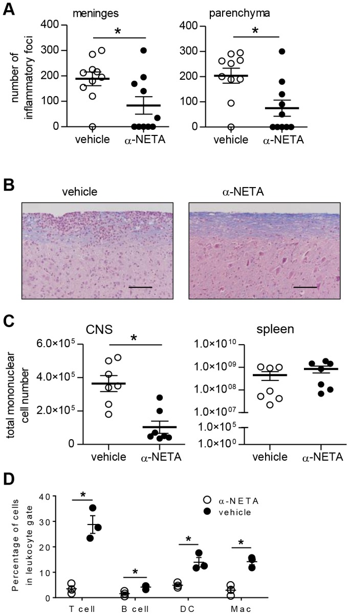
Figure 4. Reduced histological EAE in α-NETA-treated mice.

(A) Brain and spinal cord tissues were harvested 16 days after active EAE induction from mice treated with α-NETA or vehicle control. Histological changes in meninges (left panel) and parenchyma (right panel) were evaluated as described in Materials and Methods. Results are pooled from two independent experiments with similar results, each with 4–6 mice per group. mean ± SEM, *p<0.05 as by Student's t test. (B) Representative spinal cord sections are shown from actively immunized vehicle- (left panel) or α-NETA- (right panel) treated mice that were euthanized at 16 days p.i. Meningeal and parenchymal mononuclear cell infiltrates typical of acute EAE in the spinal cord of a vehicle-treated mouse (left panel). Less meningeal and parenchymal infiltration, as well as reduced myelin loss (indicated by Luxol fast blue staining intensity), in the spinal cord of α-NETA-treated mice with EAE (right panel). Bar, 50 µm. (C) At day 16 p.i., mononuclear cells isolated from the CNS and spleen of EAE mice treated with α-NETA or vehicle control were enumerated by flow cytometry. n = 7 mice/group; mean ± SEM; *p<0.05 by Student's t test. (D) CNS mononuclear cells were isolated at day 16 p.i. from EAE mice treated with α-NETA or vehicle control. Cells were then stained with fluorophore-labeled monoclonal antibodies to identify the indicated leukocyte subsets: T cells (CD3+), B cells (B220+), dendritic cells (DC; CD3-B220-NK1.1-CD11c+) and macrophages (Mac; F4/80+CD11b+). *p<0.05 by Student's t test. Two independent experiments yielded similar results.
