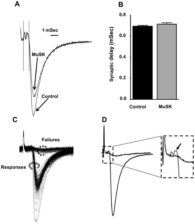
Figure 8. Reduced motor-nerve terminal excitability, but not prolonged synaptic delay, is responsible for high-frequency EPC failures of MuSK-MG-affected NMJs.
(A) Representative extracellular recordings at NMJ of TS preparations isolated from control and MuSK-MG-affected mice lacking EPC failures. Each trace is the average of 120 recordings acquired at 50 Hz. Synaptic delay was measured as the time from the beginning of the nerve terminal current to the time at which the EPC amplitude was 10% of its maximum value, as indicated by the vertical lines following the stimulus artifact. (B) Synaptic delay is equivalent for control (70 NMJs, 4 mice) and MuSK-MG-affected (60 NMJs, 4 mice) muscles. (C) Series of 120 extracellular recordings made at an endplate of a MuSK-MG-affected NMJ, which exhibited partial failure in response to 50 Hz nerve stimulation. Double circled records clearly possessed EPCs, while dot circled records were failures of EPC initiation. (D) Average of double-circled and dot circled records of panel C show that failures of EPC initiation resulted from lack of nerve impulse entry into the nerve terminal; the boxed portion of the averaged records is amplified in the inset with an arrow indicating nerve terminal current in association with the EPC.

