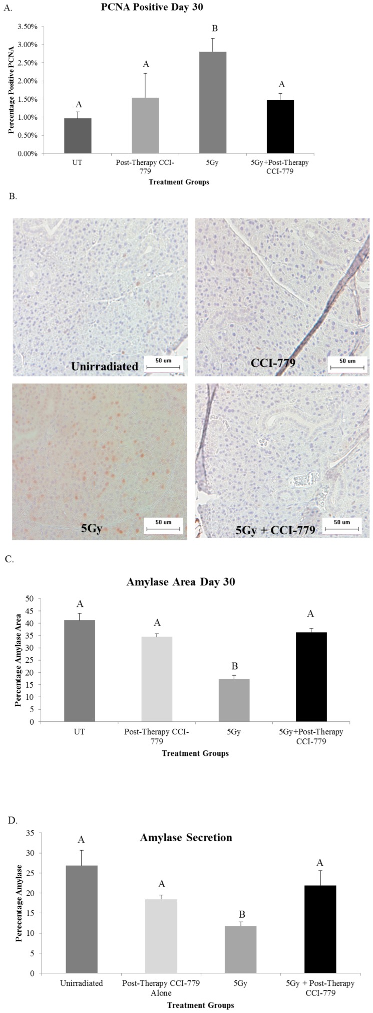
Figure 6. Radiation plus post-therapy CCI-779 improves parotid acinar cell proliferation indices and amylase production levels similar to unirradiated mice.
The head and neck region of FVB mice was exposed to a single 5-Gy dose of radiation and mice received injections of vehicle or CCI-779 on days 4-8 following initial radiation treatment. Parotid salivary glands were then collected 30 days following treatment. Significant differences (p<0.05) were determined using an ANOVA followed by a post-hoc Bonferroni multiple-comparison test. Letters above treatment groups are used to signify statistical significance; treatment groups with the same letters are not significantly different from each other. Data are presented as the mean ±SEM. UT: unirradiated. A.) Serial sections were stained for PCNA, a marker of proliferation, and the graph represents the number of acinar cells with positive PCNA staining in the parotid glands as a percentage of the total number of acinar cells. n = 4 per genotype/per treatment. B.) Representative images of positive PCNA staining. C.) Serial sections were stained to determine positive amylase area of the parotid glands. The graph represents the positive amylase area as a percentage of the parotid area as a whole. n = 4 per genotype/per treatment. D.) Stimulated saliva was collected from mice 30 days after treatment with a single 5Gy dose of targeted head and neck radiation and analyzed for total protein content as described in Materials and Methods. The graph represents the percentage of amylase protein (ranging from 50–57 kD); n = 10 per genotype/per treatment.

