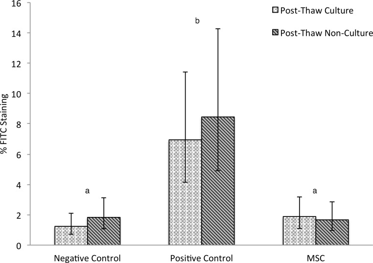
Figure 1. Mean FITC anti-BrdU staining following two-way MLR.
Lymphocyte proliferation of un-stimulated (negative control) and allogeneic stimulated (positive control) compared to stimulated lymphocytes treated with MSC. Three biological replicates and three technical replicates were used in the negative control, positive control, and for evaluation of each of the five MSC cultures. Five thousand lymphocytes in each sample were observed and designated either FITC positive (proliferative in the previous 24 h) or FITC negative (not proliferative) Umbilical cord blood MSC from five cultures were either added after a 5-day post-thaw-culture period or identical MSC vials were thawed immediately prior to the experiment. No difference was observed between post-thaw-culture and post-thaw-non-culture groups. Different letters indicate statistically significant differences between means (α = 0.05). Error bars represent the 95% confidence interval.

