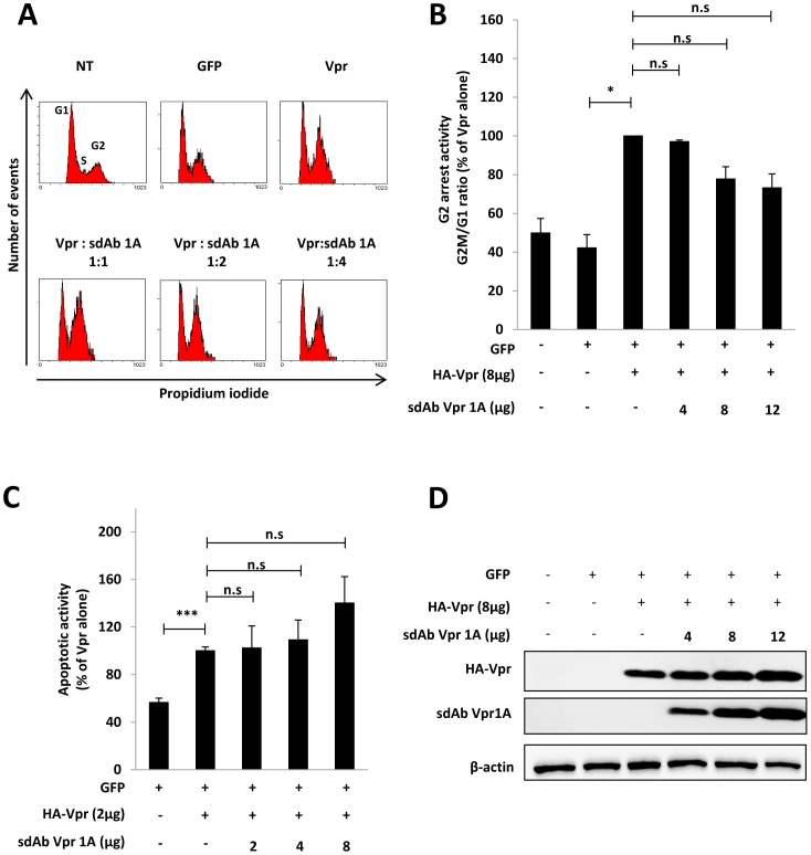
Figure 5. Impact of sdAb-Vpr1A on the G2-arrest and pro-apoptotic activities of Vpr.
HeLa cells were co-transfected with plasmids for expression of GFP and HA-Vpr in combination with increasing concentrations of c-myc tagged sdAb Vpr1A expression plasmid when indicated. (A, B) Cell cycle analysis. 48 h after transfection, cells were fixed, permeabilized, and stained with propidium iodide. The DNA content was analyzed by flow cytometry on GFP-positive cells. In A, the cell DNA content profiles from a representative experiment are shown. The cells in G1, S and G2/M phases are indicated on the upper right panel. In B, results are expressed as the percentage of the G2M/G1 ratio relative to that measured in cells expressing HA-Vpr alone and are the means of 3 independent experiments. Error bars represent 1 S.D. from the mean. Statistical significance was determined using students t test (n.s., p>0.05; *, p<0.05). C) Pro-apoptotic activity. 72 h after transfection, cell surface PS exposure was analyzed by flow cytometry on GFP positive cells after staining with phycoerythrin-labelled Annexin V and 7AAD (7-Aminoactinomycin). Results are expressed as the percentage of GFP-positive cells displaying surface PS exposure = relative to cells expressing HA-Vpr alone, and are the means of 3 independent experiments. Error bars represent 1 S.D. from the mean. Statistical significance was determined using students t test (n.s., p>0.05; *, p<0.05; **, p<0.01;***, p<0.001). D) Expression of Vpr and sdAb Vpr1A proteins. Lysates from HeLa transfected cells were analyzed by Western blotting using anti-HA (upper panel), anti-c-myc (middle panel) and anti-β-actin antibodies.

