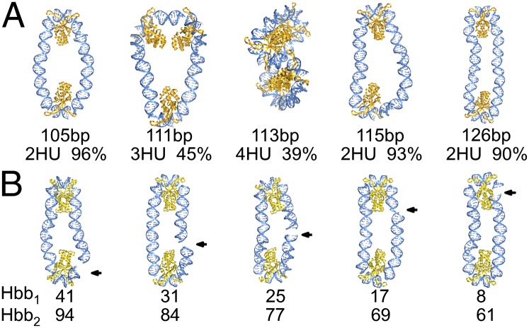
Fig. 2.
Representative molecular images, rendered in PyMOL (www.pymol.org), illustrating (A) the dominant spacings of HU on DNA minicircles of different lengths and (B) the variable sites of covalent chain closure, noted by arrows, relative to the positions of protein on 105-bp Hbb-decorated rings. Structures are aligned such that the longest principal axes are vertical, and, except for the 113-bp minicircle bearing four HU dimers, the shortest axes are perpendicular to the plane of the page. Chain lengths and populations with the depicted number of HU molecules and sequential positions of Hbb centers are denoted below the respective images. DNA is shown as a blue ribbon with attached bases, HU as a dark-gold ribbon, and Hbb as a light-gold ribbon.

