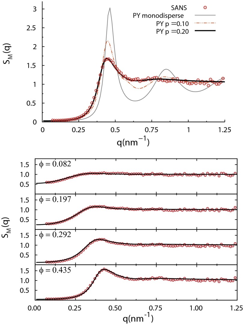
Fig. 3.
Comparison of experimental structure factors for α-crystallin solutions with polydisperse hard-sphere Percus–Yevick liquid-structure models, showing the basis for the parameter values and nm used in the present work. (Top) SAXS for mg/mL, corresponding to a deduced . Percus–Yevick predictions are shown for three polydispersity parameters: (monodisperse, thin gray line), (dotted-dashed red line), and (thick black line). closely reproduces the measured , whereas the less polydisperse and monodisperse models do not match the data. (Bottom) Over the entire range of lower α-crystallin concentrations measured, the same polydisperse Percus–Yevick with (solid lines) closely reproduces the experimental .

