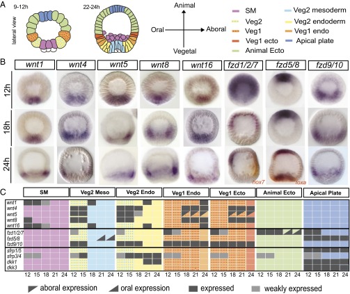
Fig. 1.
Spatial expression of Wnt-signaling genes. (A) Schematic representation of early developmental stages of S. purpuratus embryos showing the spatial arrangement of regulatory-state domains. SM, skeletogenic mesoderm; “veg1” and “veg2” denote cell lineages descended from the sixth cleavage ring of eight sister cells, each giving rise to the parts of the embryo indicated in the diagrams; “veg2 mesoderm” is also known as “nonskeletogenic mesoderm”; “animal ectoderm” and “veg1” ectoderm denote both oral and aboral ectodermal domains. (B) WMISH of significantly expressed wnt and frizzled genes at selected time points (12 h, 18 h, and 24 h); additional time points for these genes and expression patterns of dkk and sfrp genes are shown in SI Appendix, Figs. S2 and S3. (C) Expression matrix for each regulatory-state domain of the examined Wnt-signaling genes, indicating whether the gen is expressed (black/gray) or not expressed (colored background) every 3 h from 12–24 h. Regulatory domains are marked by the color code used in A. Developmental stages include 12 h (early blastula), 15 h (midblastula), 18 h (hatching blastula), and 24 h (mesenchyme blastula).

