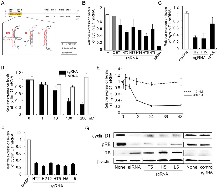
Figure 1. Design of an sgRNA targeting human cyclin D1 mRNA and the reduction in cyclin D1 mRNA and protein level by sgRNA.
(A) Design of an sgRNA targeting human cyclin D1 mRNA. Secondary structures of sgRNA/target complexes between the sgRNA and the human cyclin D1 mRNA. Arrows indicate the expected tRNase ZL cleavage sites, with numbers at the cleavage sites from GenBank data (#NM_053056.2). (B and C) Reduction in cyclin D1 mRNA level following sgRNA transfection. HSC-2 cells were plated at 1×10⁵ cells/cm2, and then cultured for 24 h. (B) sgHT1–6 (200 nM), siRNA (10 nM), sgLucHep3 (C) (10 nM) or vehicle (-) was transfected with transfection reagent; (C) sgHT2 or sgHT5 (200 nM each), siRNA (10 nM) or sgLucHep3 (control) was treated without transfection reagent after which the cells were cultured for a further 24 h. Total RNA was extracted from the cells and the cyclin D1 mRNA level was determined by qRT-PCR. (D) Dose-dependent regulation of cyclin D1 mRNA expression. HSC-3 cells were plated and cultured as above. After 24 h, naked sgHT2 or siRNA was added at the indicated concentration. (E) Time-dependent regulation of cyclin D1 mRNA expression. HSC-3 cells were plated and cultured as above. After 24 h, naked sgHT2 was added at 0 (DASHED LINE) OR 200 nM (SOLID LINE) (E), and the cells were cultured for the indicated period. Cyclin D1 mRNA level was determined by qRT-PCR. (F) Effect of sgRNA subtype on the reduction of cyclin D1 mRNA expression. HSC-3 cells were plated and cultured as above. After 24 h, naked sgHT2, sgH2, sgL2, sgHT5, sgH5, sgL5 or sgLucHep2 (control) was added at 200 nM, after which the cells were cultured for 24 h. Cyclin D1 mRNA level was determined by qRT-PCR. (G) Regulation of the cyclin D1, RB and pRB protein level by sgRNA using western blot analyses. HSC-3 cells were plated and cultured as above. After 24 h, naked sgHT2, sgHT5, sgL5, sgLucHep2 (control) (200 nM each) or siRNA (20 nM) was added using no transfection reagent (sgRNA) or with Lipofectamine 2000 (siRNA), after which the cells were cultured for 24 h. The levels of cyclin D1, RB and pRB protein in the cells were determined by western blot analysis using appropriate antibodies. Blots were re-probed for β-actin as control. Each assay represents a separate experiment performed in triplicate. Data are presented as means ± S.D.

