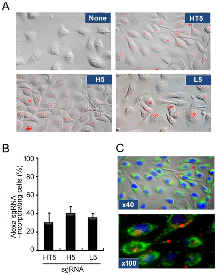
Figure 2. Confocal microscopic analysis for uptake and intracellular localization of sgRNA.
(A) HSC-3 cells were plated and cultured for 24 h. Naked Alexa568-3′-labeled sgHT2, sgH5 or sgL5 was added at 200 nM and then cells were cultured for a further 24 h, after which the cells were observed by confocal microscopy as described in Materials and Methods. (B) Red fluorescent cells were counted under a fluorescence microscope and the percentage of stained cells was calculated. Each assay represents a separate experiment performed in triplicate. Data are presented as means ± S.D. (C) HSC-3 cells were treated with naked Alexa568-3′-labeled sgHT5 and then labeled with Mitotracker Green and Hoechst33342 to visualize mitochondria and nuclei, respectively. Cells were observed through a 40x objective lens (upper panel) or 100x objective lens (lower panel) of the microscope.

