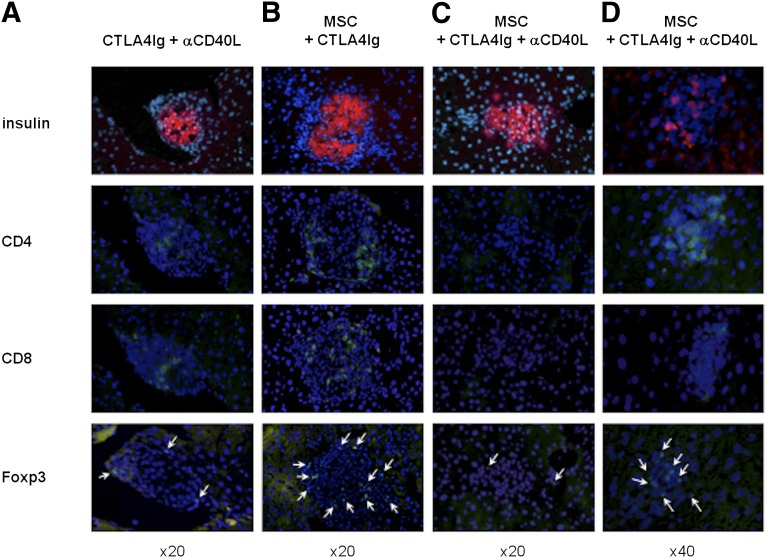
Figure 2.
Islet histological findings. Histological evaluation of serial sections of islet grafts in the liver on postoperative day 100. Immunofluorescent staining for insulin (red), Foxp3, CD4, and CD8 (green) was performed on recipient livers treated with (A): CTLA4Ig plus anti-CD40L, (B): MSCs plus CTLA4Ig, and (C, D): MSCs plus CTLA4Ig plus anti-CD40L. 4′6-Diamidino-2-phenylindole (blue) was used for nuclear staining. Foxp3 stained cells are indicated by arrows. Original magnification, ×20 (A–C), ×40 (D). Abbreviations: αCD40L, anti-CD40L; MSC, mesenchymal stromal cell.

