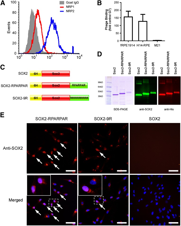
Figure 3.
Preparation and characterization of SOX2 recombinant proteins. (A): Flow cytometry analysis of the expression of NRP1 and NRP2 in human fRPE1914 cells. (B): Binding of RPARPAR phage to RPE cells. The M21 cell line, which does not express NRP1 or NRP2, was used as a negative control. The results are normalized by the phage expression levels of seven glycines. (C): Schematic of three SOX2 recombinant proteins. (D): Purified recombinant proteins were characterized using SDS-PAGE, Coomassie Blue staining, and immunoblotting. (E): Representative images of human fRPE1914 cells internalizing recombinant proteins fused with different peptides after 16 hours of incubation. Arrows: nucleolus-localized SOX2 protein. Scale bars = 50 μm. Abbreviations: fRPE, fetal retinal pigmented epithelial cells; G7, seven glycines; NRP1, neuropilin-1; NRP2, neuropilin-2; SDS-PAGE, sodium dodecyl sulfate polyacrylamide gel electrophoresis.

