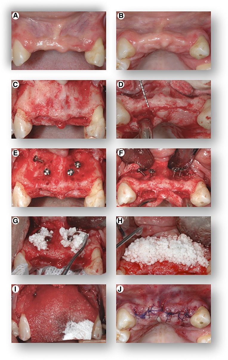
Figure 3.
Cell transplantation procedure. Front view (A) and top view (B) of the initial clinical presentation showing severe hard and soft tissue alveolar ridge defects of the upper jaw. Following elevation of a full-thickness gingival flap, the images show front view (C) and top view (D) of the severely deficient alveolar ridge, clinically measuring a width of only 2–4 mm. Front view (E) and top view (F) of the placement of “tenting” screws in preparation of the bony site to receive the graft. Placement of the β-tricalcium phosphate (seeded with the cells 30 minutes prior to placement at room temperature) into the defect (G), with additional application of the cell suspension following placement of the graft in the recipient site (H). Placement of a resorbable barrier membrane (I) to stabilize and contain the graft within the recipient site, and top view (J) of primary closure of the flap.

