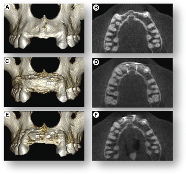
Figure 4.
Cone-beam computed tomography (CBCT) scans. CBCT scans were used to render three-dimensional reconstructions of the anterior segment of the upper jaw and cross-sectional (top view) radiographic images to show volumetric changes of the upper jaw at three time points. (A, B): The initial clinical presentation shows 75% jawbone width deficiency. (C, D): Immediately following cell therapy grafting, there is full restoration of jawbone width. (E, F): Images show 25% resorption of graft at 4 months and overall net 80% regeneration of the original ridge-width deficiency.

