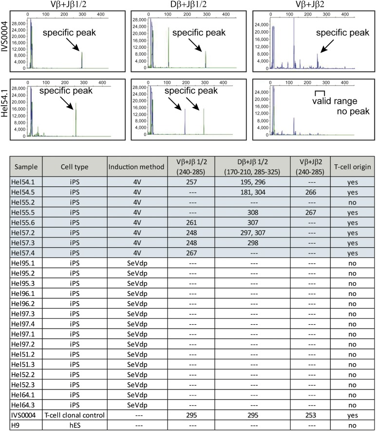
Figure 4.
Origin of iPSCs. Multiplex polymerase chain reaction fragment size analysis of T-cell receptor β region was performed by capillary electrophoresis using ABI3730 DNA analyzer. Specific peak profiles are shown for positive clonal T-cell control (IVS0004) and for one representative iPSC line (HEL54.1) produced by the 4V method. Fragment sizes for all iPSC clones are indicated in the table. DNA from hES cells (H9) was used as a negative control for the experiments. Abbreviations: 4V, Sendai virus (Cytotune); hES, human embryonic stem cell; iPSC, induced pluripotent stem cell; SeVdp, tetracistronic Sendai virus.

