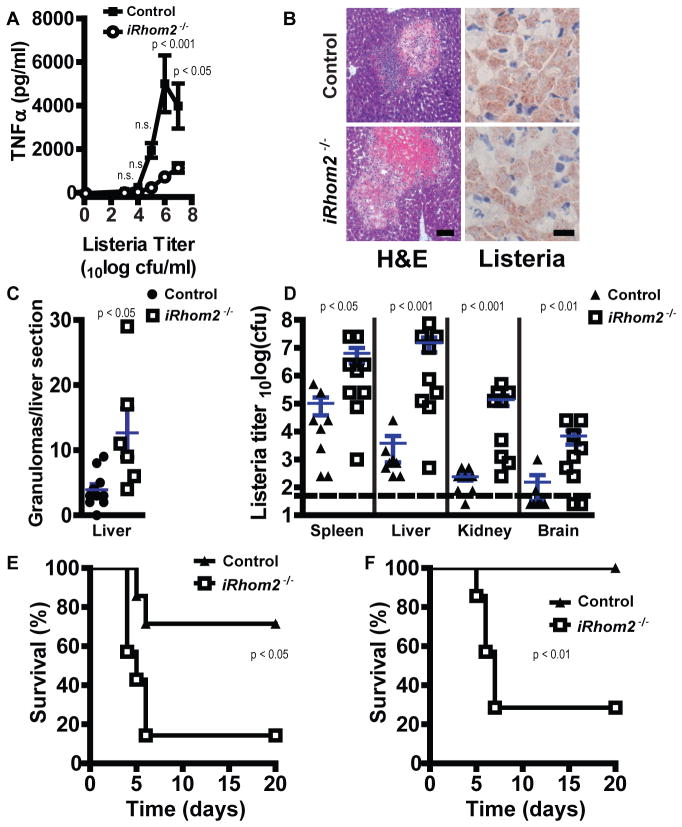
Fig. 4.
iRhom2 is crucial for control of L. monocytogenes. (A) TGEMs (105) isolated from control or iRhom2−/− mice (n = 5 to 8 per group) were exposed to the indicated titers of L. monocytogenes for 24 hours. TNFα in culture supernatants was determined by ELISA (means ± SEM). (B) Control and iRhom2−/− mice (n = 6 to 10 per group) were infected with 104 colony-forming units (cfu) L. monocytogenes. Livers were isolated on day 4 after infection, sectioned, and stained with H&E (left) or with antibody against Listeria (right). Scale bars: left, 100 μm; right, 20 μm. (C) Granulomas were counted in F4/80-stained liver sections (not shown) from the mice in (B). Data points are granulomas or liver of individual mice. Blue lines, means ± SEM (n = 6 to 10 per group). (D) Control and iRhom2−/− mice (n = 8 to 9 mice per group) were infected with 105 cfu L. monocytogenes, and bacterial titers were determined in spleen, liver, kidney, and brain on day 4 after infection. Data points are titers of individual mice. Dashed line, limit of detection. Blue lines, means ± SEM. (E and F) Control and iRhom2−/− mice (n = 7 per group) were infected with 5 × 10⁴ cfu (E) or 5 × 10³ cfu (F) L. monocytogenes, and mouse survival was monitored for 20 days.

