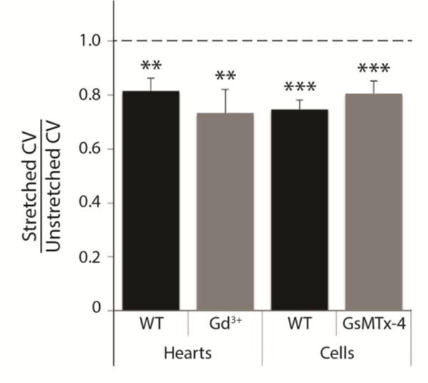
Figure 2.

Conduction slowing with stretch is not affected by stretch-activated channel blockade in isolated hearts or myocytes. Mean conduction velocity in mechanically loaded hearts and cells was 70–80% of unstretched values, and significantly different from the original value (1.0), in the following four conditions: untreated hearts (N=5, P<0.01), hearts with stretch-activated channel blockade by Gd3+ (N=5, P<0.01), untreated cells (N=8, P<0.001), and cells with stretch-activated channel blockade by peptide GsMTx-4 (N=3, P<0.001 cells). There was no significant difference in the CV ratio between control (WT) groups and treated groups for hearts or cells.
