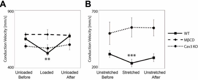
Figure 3.

Stretch-dependent conduction slowing requires caveolae in isolated mouse hearts (A) and cultured myoctes (B). (A) Conduction slowing observed with loading in WT hearts is not seen when caveolae are depleted via either treatment with MβCD or genetic deletion of caveolin-3 (**WT hearts N=5, P<0.01; MβCD-treated and Cav3 KO hearts N=5, P=N.S.). (B) Conduction slowing observed in WT cells is prevented when caveolae are depleted via genetic deletion of caveolin-3 (***WT cells N=8 P<0.001; Cav3 KO cells N=3, P=N.S.). Within paired observations, only untreated WT hearts and myocytes show statistically significant changes with stretch.
