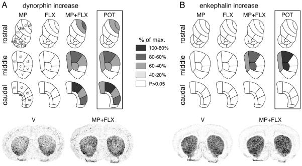
Figure 1.
Topography of potentiated neuropeptide expression in the striatum after repeated methylphenidate plus fluoxetine treatment. Maps depict the distribution of the increases (vs. V) in dynorphin (A) and enkephalin expression (B) in the rostral, middle and caudal striatum after 5 daily injections of methylphenidate (5 mg/kg, i.p.; MP), fluoxetine (5 mg/kg; FLX) or methylphenidate+fluoxetine (5 mg/kg each; MP+FLX). Potentiation (POT) denotes the difference between methylphenidate+fluoxetine and methylphenidate groups. The increases are expressed relative to the maximal increase for each neuropeptide (% of max.). Sectors with significant differences [vs. vehicle (V) controls, or methylphenidate+fluoxetine vs. methylphenidate (POT)] (P<0.05) are coded as indicated. Sectors without significant effects are in white. Illustrations of film autoradiograms depicting the expression of dynorphin (left) and enkephalin (right) in coronal sections from the middle striatum after repeated treatment with vehicle (V) or methylphenidate+fluoxetine (MP+FLX) are shown below the maps. Abbreviations: caudate-putamen: c, central; d, dorsal*; dc, dorsal central; dl, dorsolateral*; dm, dorsomedial; m, medial; v, ventral; vc, ventral central; vl, ventrolateral*; nucleus accumbens: mC, medial core; lC, lateral core; mS, medial shell; vS, ventral shell; lS, lateral shell; *sensorimotor sectors (see Yano and Steiner, 2005a).

