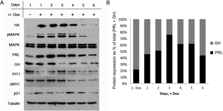
Figure 2.
Stable expression of V12Ras increases PRL and reduces GH expression. A, Western blot analysis of GH4 clone number 10 treated ±dox. GH4 clone number 10 cells expressing dox-inducible 3HA-V12Ras (from Figure 1) were serum starved (0.05% FBS) overnight. Serum-starved medium containing 2 μg/mL dox was added on days 1, 3, and 5, and cells were harvested daily for 6 days. For day 1, (−)Dox cells were harvested after overnight serum starvation and were not treated with dox. Whole-cell extracts (50 μg) were separated by SDS-PAGE and probed with the antibodies listed (n = 3 independent experiments; a representative experiment is shown). B, Quantification of PRL and GH expression ±dox. PRL (black bars) and GH (gray bars) protein expression from panel A was quantified by densitometric analysis using AlphaImager software (Alpha Innotech), normalized to PRL expression on day 1 (−)Dox, and graphed as a percentage of total (PRL + GH) expression for each day.

