Figure 3.
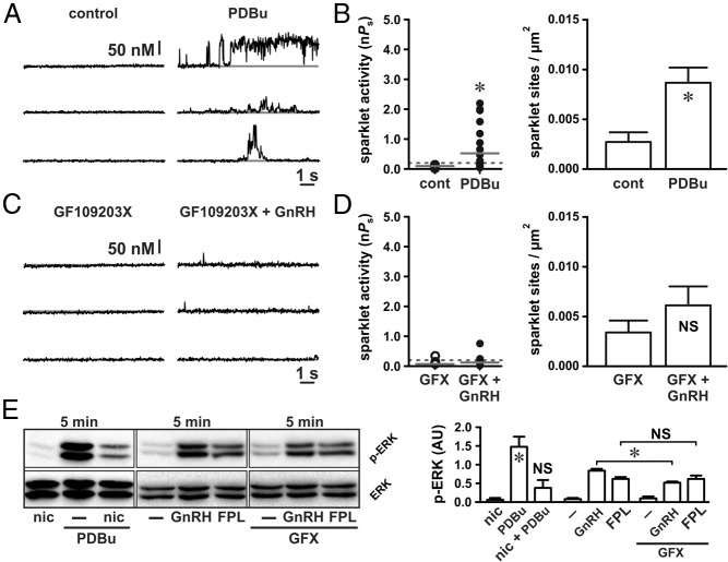
GnRH-induced L-type Ca2+ channel sparklets and ERK activation require PKC. A and C, Representative traces showing time courses of Ca2+ influx in an αT3–1 cell before and after application of the PKC activator PDBu (50 nM) (A) and GnRH (3 nM) (C) in the presence of the broad-spectrum PKC inhibitor GFX (1 μM). B and D, Plots of Ca2+ sparklet site activities (nPs) and mean ± SEM Ca2+ sparklet site densities (Ca2+ sparklet sites per square micrometer) before and after PDBu (n = 11 cells) (B) and GnRH in the presence of GFX (n = 9 cells) (D). E, Western blot analysis of ERK activation (as measured by ERK phosphorylation) in αT3–1 cells exposed to PDBu (50 nM) or PDBu with nicardipine (1 μM) for 5 minutes and αT3–1 cells exposed to GnRH (3 nM) or FPL 64176 (500 nM) for 5 minutes in the presence or absence of GFX (1 μM; n ≥ 3 independent experiments). *, P < .05. cont, control; nic, nicardipine.

