
Figure 4.
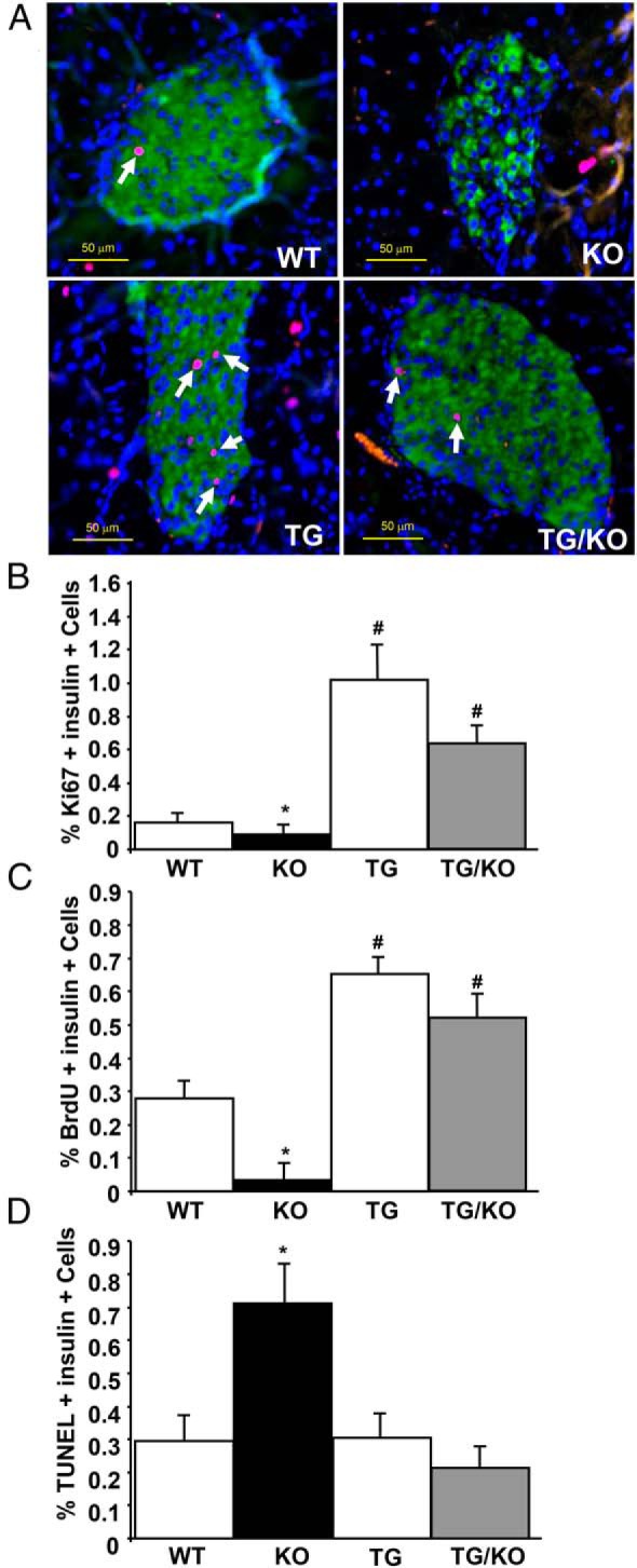
β-Cell proliferation and survival in RIP-HGF TG mice with IRS2 deficiency. A, Representative photomicrographs of mouse pancreatic sections stained for insulin (green), DAPI (blue), and Ki67 (red) from WT, TG, KO, and TG/KO mice at 6 weeks of age. Arrows indicate Ki67-positive β-cell nuclei. B and C, Quantification of the percentage of Ki67-positive (B) and BrdU-positive (C) β-cells. D, TUNEL-positive β-cells to detect cell death in WT (n = 7), TG (n = 6), KO (n = 7), and TG/KO (n = 9) mice. Results are means ± SEM. *, P < .05 vs the other 3 groups of mice; #, P < .05 vs WT.