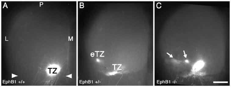
Figure 2. EphB1 is required for appropriate retinocollicular mapping.

(A) Dorsal view of the superior colliculus (SC) of a WT mouse at P8, one day after injection of DiI into temporal retina. A dense, focal termination zone (TZ) in the appropriate location in anterior (arrowheads) SC is evident. (B and C) The SC of EphB1+/- and EphB1-/- mice one day after an injection of DiI similar to that in (A). (B) In 39% of EphB1+/- cases, ectopic termination zones (eTZs) of temporal RGC axons are evident lateral (L) to a normal appearing TZ. (C) Similarly, in EphB1-/- mice, 48% of cases have an eTZ positioned laterally. Scale bar=500μm. M, medial; P, posterior.
