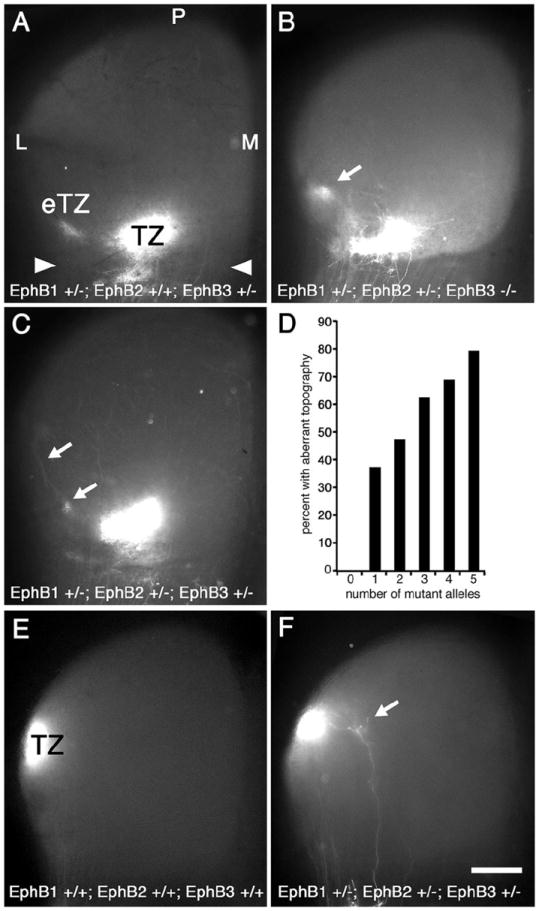
Figure 3. Number of EphB1, EphB2, and EphB3 null alleles corresponds to frequency, but not severity, of aberrant mapping phenotype.

Dorsal view of the superior colliculus (SC; anterior SC marked by arrowheads) of P8 mice injected with DiI in temporal retina one day earlier. Mice deficient for one or more alleles of EphB1, EphB2, and/or EphB3 have retinotopic defects. (A) EphB1+/-; EphB2+/+; EphB3+/- mice have an appropriately positioned termination zone (TZ) as well as an ectopic TZ (eTZ, arrows) lateral (L) to the main TZ. Qualitatively similar defects are observed in (B) EphB1+/-; EphB2+/-; EphB3-/- mice and in (C) EphB1+/-; EphB2+/-; EphB3-+/- mice. (D) Graph correlating the number of combined mutant EphB1, EphB2, and EphB3 alleles to the presence of an aberrant retinotopic map. Each additional missing allele increases the frequency of an aberrant map. For 0-5 missing EphB alleles, n=10, 48, 36, 32, 32, and 24, respectively (right, 182 total cases with temporal DiI injections). Fisher’s exact test indicates statistical significance at p<0.03 for one missing EphB allele, p<0.01 for two and three missing alleles, p<0.001 for four and five missing alleles. (E) SC from a wild type mouse after injection of DiI in dorsal retina showing a dense TZ in the appropriate position in lateral SC (n=6). (E) DiI injection in dorsal retina of an EphB1+/-; EphB2+/-; EphB3-+/- mouse reveals, in addition to a normal TZ, a small eTZ positioned medially (arrow) in some cases (n=7). Scale bar=500μm. M, medial; P, posterior.
