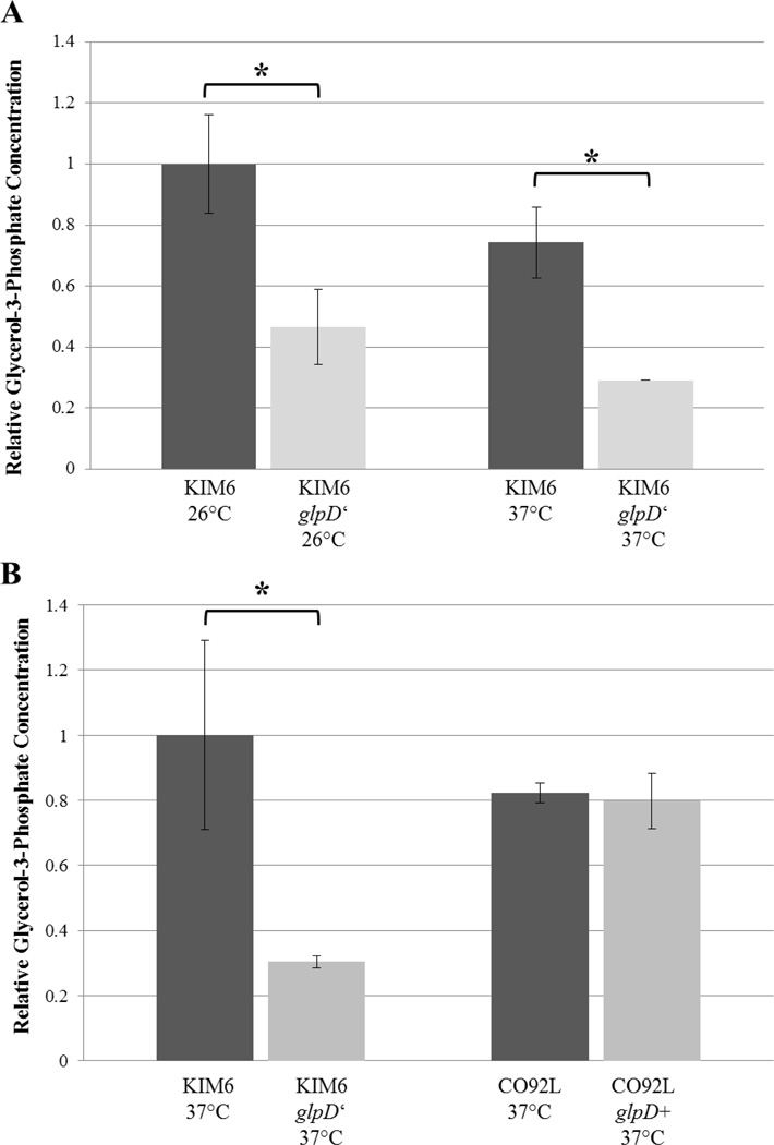
FIG. 4.
Colorimetric glycerol-3-phosphate determination (Biovision) of glpD allelic exchange mutants. A. KIM6 and KIM6 glpD’ were grown to logarithmic phase in HIB media supplemented with 0.2% glycerol at both 26°C and 37°C. Approximately 2×10⁹ total cells as determined by optical density were pelleted, lysed, and processed by methanol/chloroform extraction. B. Intracellular concentrations of glycerol-3-phosphate quantification of KIM6, KIM6 glpD’, CO92L, and CO92L glpD+ direct lysates of 2×10⁹ 37°C logarithmic phase cultures. Relative glycerol-3-phosphate concentrations were determined from 50 µl, 25 µl, 12.5 µl aqueous phase extract (A) or cellular homogenate supernatant (B) per sample. Error bars reflect standard deviation from the mean. * P-value < 0.05 as determined by Student’s T test.

