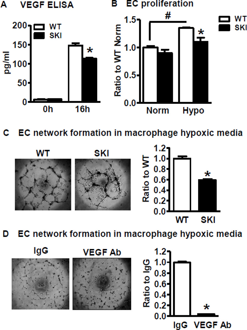
Figure 6. SERCA 2 C674 regulated VEGF production by hypoxic macrophages induces endothelial cell angiogenic function.
A. VEGF concentration measured by ELISA in the culture media of WT or SKI macrophages exposed to hypoxia for 16 h. *P<0.05, vs. WT, n=3. B. Proliferation assay of ECs exposed to conditioned media of hypoxic macrophages. ECs were seeded into 96-well plates and treated with media from WT or SKI macrophages exposed to normoxia (Norm) or hypoxia (Hypo) for 72 h. Proliferation of ECs was analyzed with water soluble tetrazolium salt assay. *p<0.05, vs. WT, n=3. C. Network formation by ECs exposed to media of hypoxic macrophages. ECs were seeded into BD Matrigel™ coated 96-well plates and treated with conditioned media from WT or SKI macrophages exposed to hypoxia for 16 h. EC network formation was determined by network length measured by Image J. *P<0.05, vs. WT, n=3. D. Effect of neutralizing anti-VEGF antibody on network formation by ECs exposed to media of hypoxic macrophages. ECs were seeded in BD Matrigel™ coated 96-well plates and treated with conditioned media from hypoxic macrophages together with rabbit IgG or VEGF neutralizing antibody (VEGF Ab). Network formation was compared after 16 h treatment. *P<0.05, vs. IgG, n=3.

