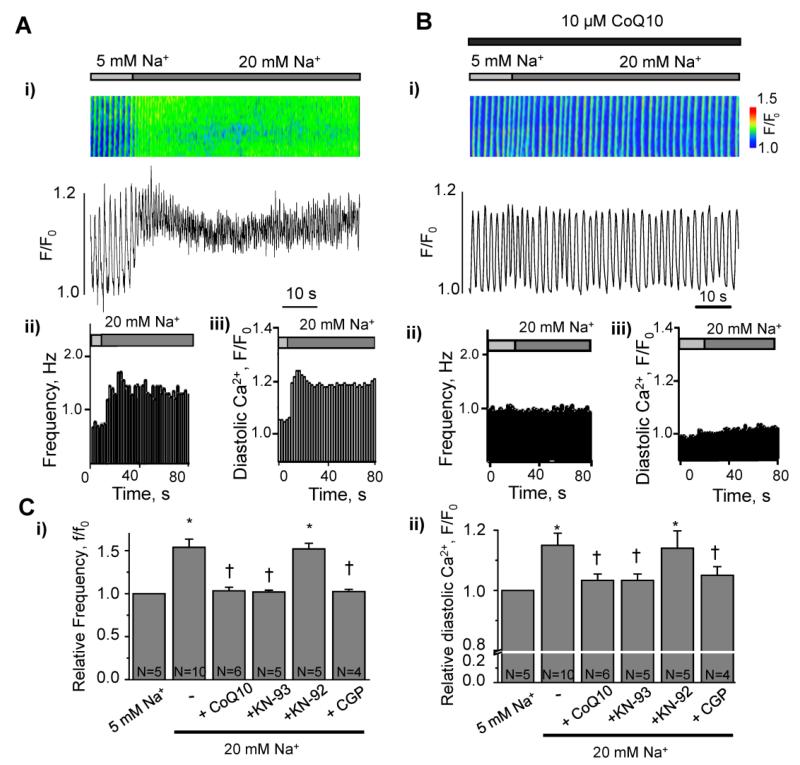
Figure 3. Role of ROS, mitochondrial Na+/Ca2+ exchange, and CaMKII in maladaptive Ca2+ handling induced by elevation of [Na+]i in membrane-permeabilized cardiomyocytes.
A. i) Confocal line-scan image and profile of Ca2+ transients in a membrane-permeabilized rabbit ventricular myocyte, recorded during elevation of cytosolic Na+ from 5 to 20 mM; ii) spontaneous Ca2+ wave frequency and iii) diastolic Ca2+ level for the experiment shown in panel (i). B. i, Confocal line-scan image and profile of Ca2+ transients in a membrane-permeabilized rabbit ventricular myocyte during elevation of cytosolic Na+ from 5 to 20 mM in the presence of the antioxidant CoQ10 (10 μM); ii) spontaneous Ca2+ wave frequency and iii) diastolic Ca2+ level for the experiment shown in (i). C. Quantification of spontaneous Ca2+ wave frequency f/f0(i) and the diastolic Ca2+ level (ii), induced by elevation of [Na+]i in the absence and presence of the antioxidat CoQ10 (10 μM), the CaMKII inhibitor KN-93 (3 μM), the inactive analog KN-92 (3 μM) or mitochondrial Na+/Ca2+ exchange inhibitor CGP-37157 (CGP, 10 (μM) N=4-10 * p<0.05 vs control; †p<0.05 vs 20 mM Na+. f0, the frequency of spontaneous Ca2+ waves at the beginning of experiment.

