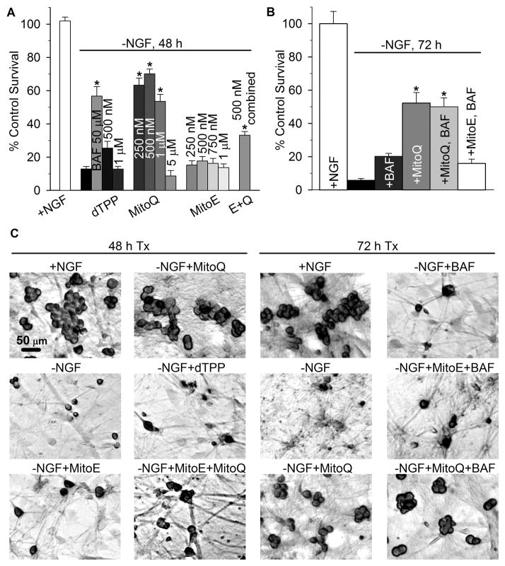
Fig. 2.
MitoQ10 prevented caspase-dependent and independent death induced by NGF deprivation. (a) To determine the effects of the mitochondria-targeted agents on apoptosis in sympathetic neurons, cultures were deprived of NGF alone or in the presence of the indicated concentrations of the broad spectrum caspase inhibitor BAF, dTPP, MitoQ10, MitoE2, or MitoE2 + MitoQ10 (E + Q, 250 nM of each). Neurons that were not committed to die by caspase- dependent death (Commitment 1) were rescued by NGF replacement at 48 h; five days later the cells were fixed, counted, and quantified as the percent of viable neurons in sibling cultures that were maintained in NGF from the time of plating. MitoQ10 treatment (0.25–1 μM) and BAF inhibited caspase-dependent death of NGF-deprived neurons (*p < 0.001). (b) To determine the effect of MitoQ10 (500 nM) and MitoE2 (500 nM) on caspase-independent death, cultures were deprived of NGF alone or with MitoQ10 or MitoE2 from the time of deprivation. These cultures were rescued with NGF 72 h later and quantified as described above. Cultures treated with BAF (50 μM) continued to die after 72 h, despite NGF readdition, indicating that the neurons were committed to die by caspase-independent death (Commitment 2). Cultures receiving MitoQ10 alone and MitoQ10 plus BAF treatment escaped impending death to a similar degree (p = 0.76), indicating that MitoQ10 treatment was neuroprotective against both caspase-dependent and-independent death after 72 h of deprivation (*p < 0.001 for MitoQ10 and MitoQ10 + BAF versus –NGF + BAF). MitoE2 had no effect on caspase- independent death. Results are presented as ±SEM from at least four independent experiments (n = 12–34 cultures). Statistical analysis was performed by ANOVA on ranks, followed by Dunns or Holm-Sidak post hoc tests for multiple comparisons between groups. (c) Representative micrographs of cultures treated as indicated, rescued with NGF after 48 or 72 h and fixed. Easily distinguishable cellular outlines of plump, crystal violet-stained neurons highlight viable neurons over the dead cells that show little or no staining.

