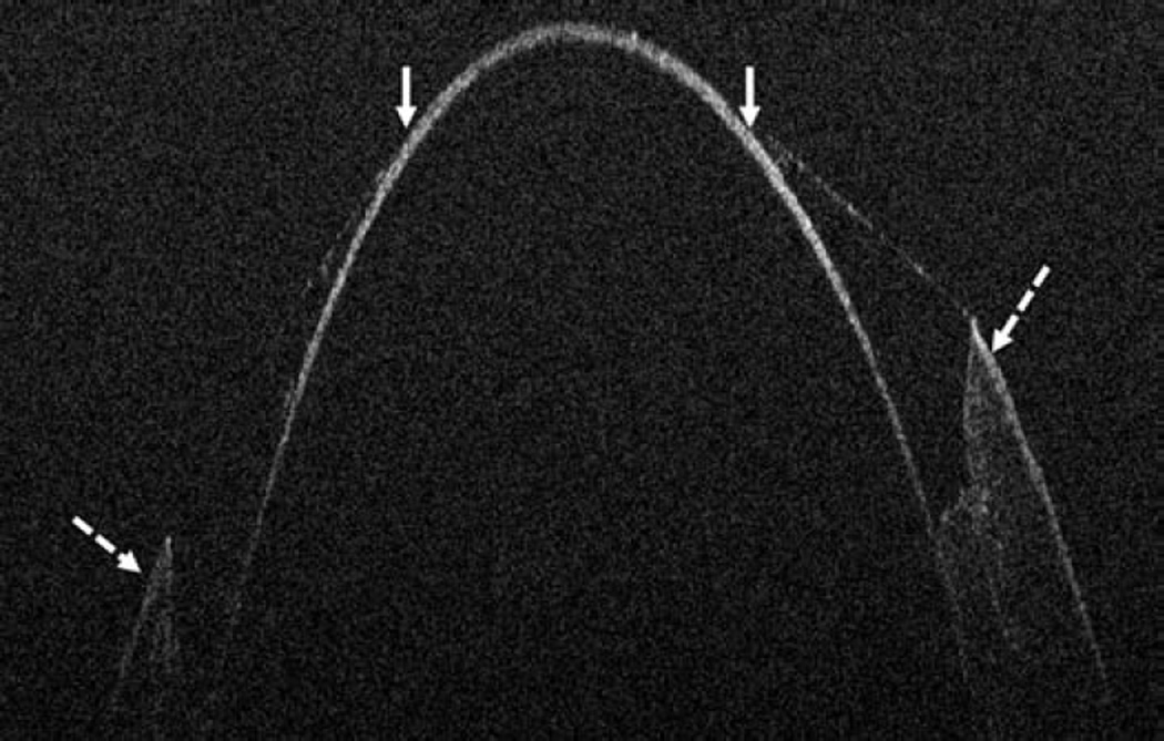
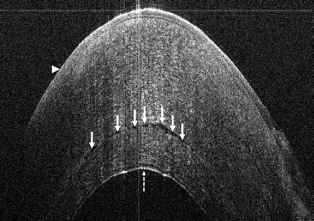
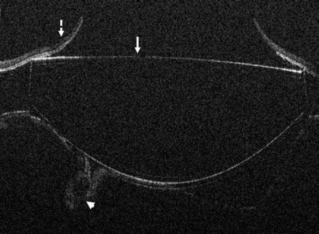
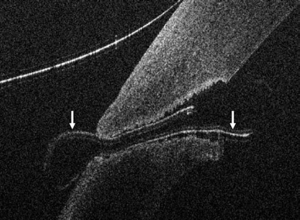
Figure 2.
Anterior segment intraoperative optical coherence tomography in lamellar keratoplasty. Intraoperative optical coherence tomography (OCT) B-scan following insertion of graft (dashed arrow) reveals partial apposition with persistent interface fluid (solid arrows) between the graft and the host cornea (arrowhead) during Descemet stripping automated endothelial keratoplasty surgery (Top). Intraoperative OCT B-scan during deep anterior lamellar keratoplasty surgery allows for visualization of edge of full-thickness cornea (dashed arrows) and bare Descemet membrane (solid arrows) following stromal dissection (Bottom).


