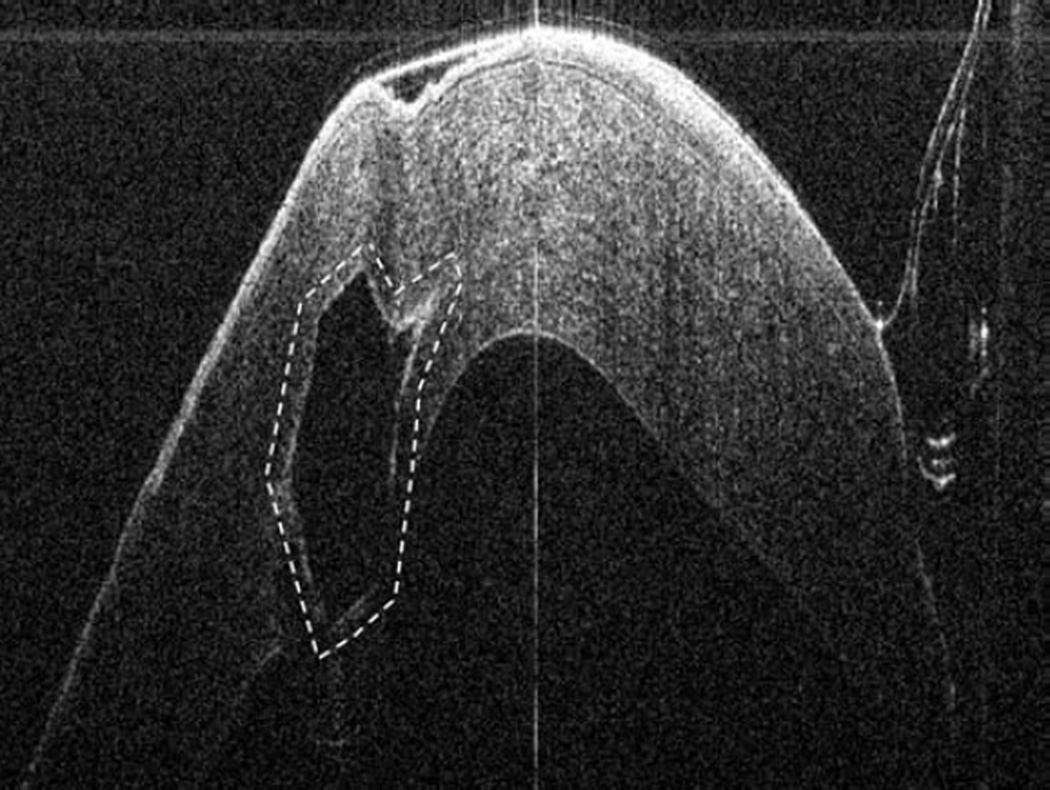
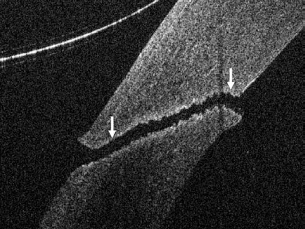
Figure 3.
Anterior segment intraoperative optical coherence tomography during corneal and cataract surgery. Intraoperative optical coherence tomography (OCT) B-scan following insertion of INTACS implant (dashed outline) confirms location (Top left). Intraoperative OCT B-scan during cataract extraction and intraocular lens placement verifies optimal location of IOL (solid arrow) behind the anterior capsule (dashed arrow). Irregularity and undulations in the posterior capsule (arrowhead) are visualized (Bottom left). Intraoperative OCT B-scan of clear corneal wound (solid arrows) with associated wound gape is visualized (Top right). Intraoperative OCT B-scan at different location of same corneal incision reveals a capsular remnant (solid arrows) within the wound resulting in wound gape (Bottom right).


