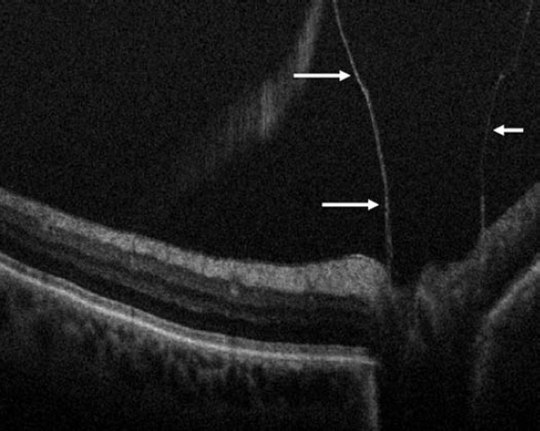
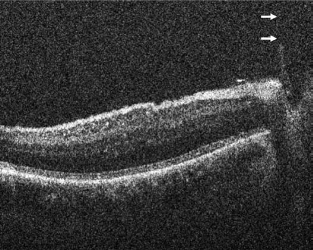
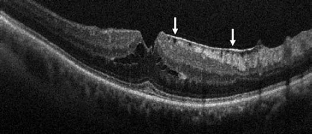
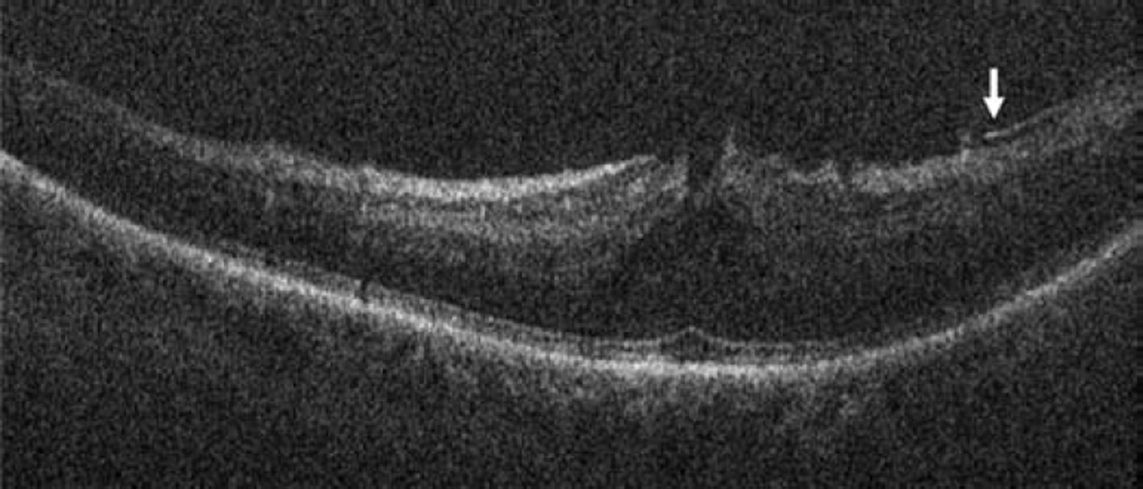
Figure 4.
Epiretinal membrane surgery and intraoperative optical coherence tomography. Preincision intraoperative optical coherence tomography (OCT) B-scan reveals epiretinal membrane (ERM, arrows, top left) and attached posterior hyaloid attached (arrows) at the optic nerve (Bottom left). Intraoperative OCT B-scan following membrane peeling and hyaloid elevation identifies residual ERM (arrow, top right) and confirms hyaloid release from the optic nerve with minimal residual hyaloid elements (arrows, bottom right).


