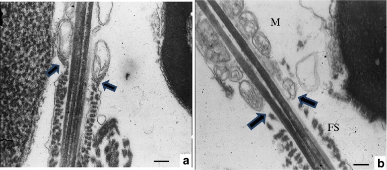
Fig. 4.
Transmission electron microscopy analysis of spermatozoa. a Normal control spermatozoon showing the annulus at the junction of the MP and the PP. b Annulus was not organized in caudal end of middle piece in spermatozoa in infertile patient diagnosed with asthenozoospermia. Note that fibrous sheath (FS) is relatively disorganized. M mitochondrial sheath. Bars: 0.25 μm

