
Fig. 2.
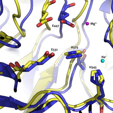
Overlay of the protein model for B. circulans rBgaD-50–650 in yellow and E.coli β-galactosidase crystal structure (PDB: 1DP0) in blue. The two catalytic glutamates (E477, E532) and two conserved histidines (H345, H397) are shown for B. circulans rBgaD-50–650. The magnesium (magenta) and sodium (light blue) ions are required for E.coli β-galactosidase, but not for the β-galactosidase of B. circulans. (For interpretation of the references to colour in this figure legend, the reader is referred to the web version of this article.)