
Fig. 6.
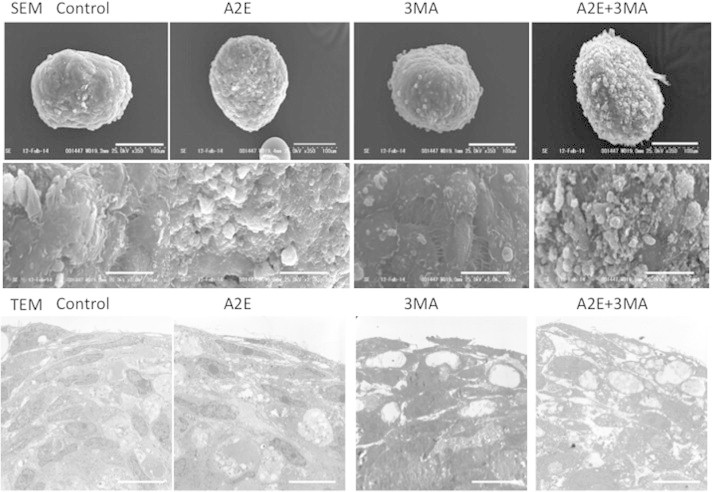
Ultrastructural analysis of human RPE spheroids. Top to bottom. SEM images (and magnified insets), and TEM images of human RPE spheroids. Human primary RPE cells were cultured in spheroids for 7 days and subjected to A2E (10 μm) and/or 3MA (10 mM) treatment. SEM revealed a smooth surface on the spheroids. With A2E treatment, budding or segmentation of RPE cells, accompanied by basal laminar deposits and membranous debris-like materials, was observed and was further pronounced in the presence of A2E and 3MA. Bars = 100 (top) and 20 (bottom) μm, respectively. TEM revealed that 3MA treatment alone resulted in infrequent vacuole formation, a markedly disorganized cellular structure of RPE with numerous vacuole-like structures, cellular materials, and increased lipid deposition in response to treatment with A2E plus 3MA. Bars = 20 μm.