


Figure 1.
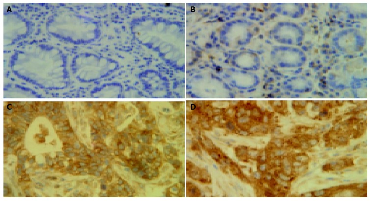
Comparison of nuclear staining rates statistically higher in tumor cells with those in adjacent and normal epithelial cells after staining of RelA. A: Normal epithelial cells with no nuclei staining; B: Adjacent epithelial cells with no nuclei staining; C: Cancer tissues with strong nuclei staining; D: Cancer metastasis to the liver with specially enhanced nuclei staining (×400).