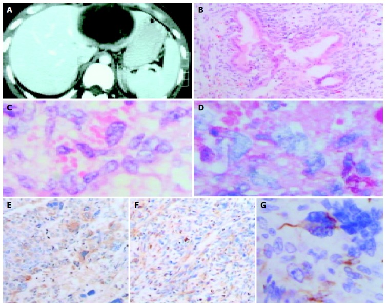
Figure 1.

The appearance of UESL in enhanced CT, H&E stain, PAS staining and immunohistochemical assay. A: Enhanced CT scan of primary tumor. It shows a well encapsulated, multiloculated cystic mass in left hepatic lobe containing internal septations and mastoid protuberances in the intracavity, and the mass projects to the anterior abdominal wall; B: Microphotograph of primary tumor showing adeniform structure surrounded by predominantly atypical, pleomorphic sarcomatous cells (H&E stain, ×100); C: Eosinophilic hyaline globules seen in the cytoplasm of some tumor cells (H&E stain, ×400); D: Eosinophilic hyaline globules showing strongly positive for periodic acid-Schiff (PAS) staining (×400); E: Immunohistochemical assay revealing spindle shaped, polygonal, multinucleated or macronuclear cells with positive staining for vimentin (SP, ×100); F: Immunohistochemical feature presenting some anaplastic tumor cells with positive staining for alpha-1-antichymotrypsin (AACT) (SP, ×100); G: Immunohistochemical assay showing many pleomorphic tumor cells with positive staining for desmin (SP, ×400).
