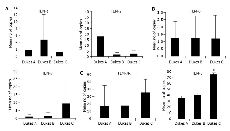
Figure 2.

Real-time quantitative RT-PCR. Shown in the figure are Mean copies/ng mRNA. Levels of expression of TEMs in colon tissues in tumors with different in different Dukes stages. The number of transcripts of TEM-1 is high in Dukes A and TEM-2 higher in Dukes B tumors. TEM-6 shows no difference in all three stages (Dukes A, B and C). Dukes C tumor expressed greatest level of TEM-8 (aP = 0.001 vs Dukes A). Both TEM-7 and -7R shows higher level of expression in Dukes C, however the difference is not significant (P>0.05).
