
Figure 4.
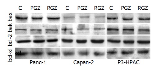
Expression of apoptotic proteins in cells treated with TZD. Sub-confluent cells were treated with 20 μmol/L of TZD (RGZ or PGZ) for 24 h. Cells were then harvested, and whole-cell protein extracts were fractionated by sodium dodecyl sulfate-polyacrylamide electrophoresis and transferred to nitrocellulose paper as described in Methods. Different proteins were detected by incubating the filter with specific antibodies.