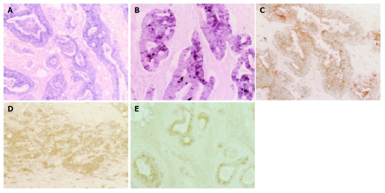
Figure 1.

Positive staining in cytoplasm of colorectal adenocarcinoma cells shown by immunohistochemical staining of Ties and Angs. Immunoalkaline phosphatase staining; Tie-1 (A), Tie-2 (B) and DAB staining; Ang-1 (C), Ang-2 (D), and Ang-4 (E). (magnification: ×100, each).
