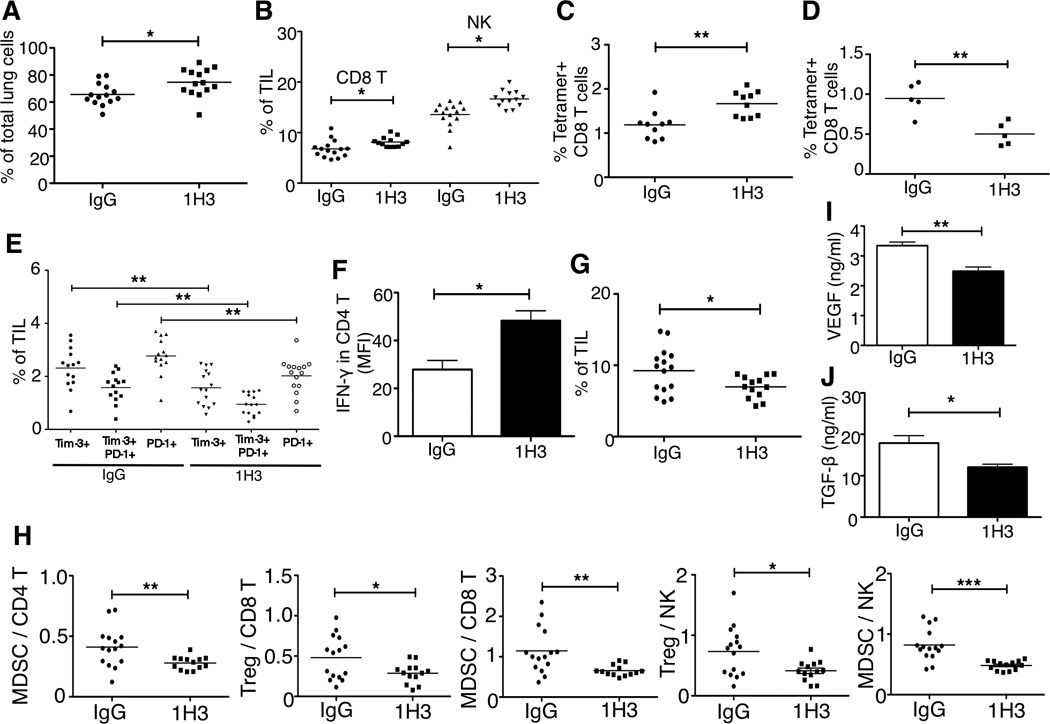
Figure 4. Anti-B7x Therapy Alters the Intratumor Balance of Anti-tumor Effector Immune Cells and Immunosuppressive Cells.

BALB/c mice were iv injected with B7x/CT26 and then treated with 1H3 or control mouse IgG. At day 17, single-cell suspensions from tumor bearing lungs were analyzed by FACS for the percentage of infiltrated CD45+ cells (A), the percentage of CD8 T cells and NK cells (B), the percentage of tumor antigen AH1 (SPSYVYHQF)-specific CD8 T cells (C), the percentage of CD4 T cells that were Tim-3+PD-1+, Tim-3+ alone and PD-1+ alone (E), and CD11b+Ly6C+ monocytic myeloid-derived suppressor cells (G). At day 17, single-cell suspensions from bloods were analyzed by FACS for the percentage of tumor antigen AH1-specific CD8+ T cells were measured (D). Cell suspensions from tumor bearing lungs were stimulated with 1x cell stimulation cocktail for 5 hours and stained with antibodies to CD3, CD4 and IFN-γ or isotype controls (F). Results are pooled from three independent experiments; *p<0.05, **p<0.01. Result of (D) is a representative data from two independent experiments. (H) The ratios of Treg (Foxp3+ CD4+) and MDSCs to CD8 T cells, CD4 T cells, and NK cells. These results are pooled from three independent experiments; *p<0.05, **p<0.01, ***p<0.001. (I) Total amount of VEGF from tumor bearing lungs was measured using ELISA. **p<0.01. (J) Total amount of TGF-β from tumor bearing lungs were measured using ELISA. Each group contained 5 mice. *p<0.05.
