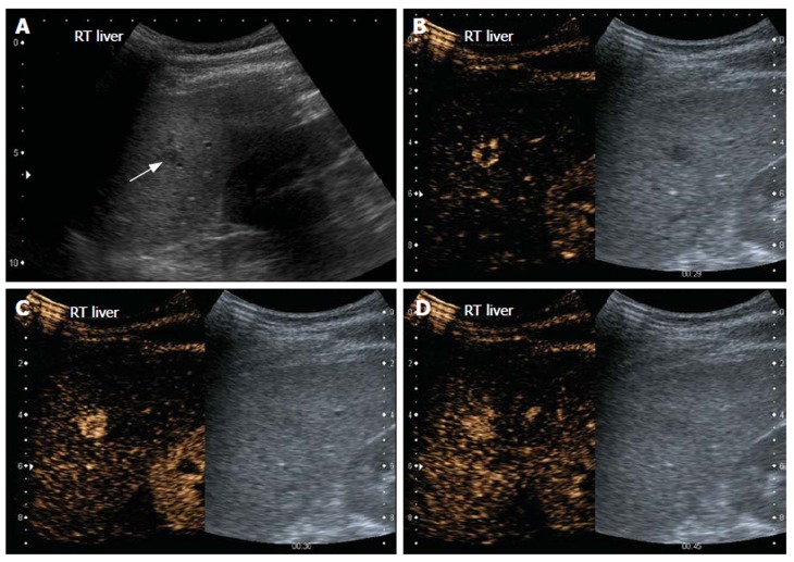
Figure 1.
Haemangioma. Gray-scale US image (A) and split-screen display images of contrast-enhanced US scan using a low MI technique (B-D). The left panel shows the contrast sensitive image while the corresponding gray-scale image is on the right. On gray-scale US, the liver is hyperechoic, consistent with fatty infiltration and an ill-defined hypoechoic lesion is seen (arrow, A). On contrast enhanced US, the lesion demonstrates peripheral nodular enhancement during the arterial phase (B). At 36 s, the lesion has almost completely filled in (C). At 45 s, the lesion is completely enhanced (D) and sustained enhancement was observed in the late phase scan (not shown).

