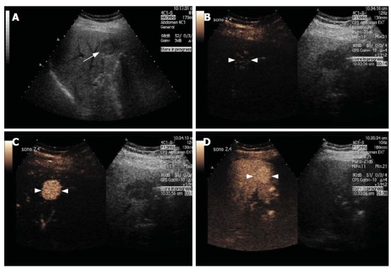
Figure 2.

Focal Nodular Hyperplasia. Gray-scale US image (A) and split-screen display images of a contrast-enhanced US scan using a low MI technique (B-D). The gray-scale US image shows a focal hypoechoic lesion (arrow, A) in a diffusely hyperechoic liver in keeping with fatty infiltration. After contrast injection, the lesion enhances avidly in the arterial phase with filling seen from a central feeding vessel, demonstrating the classical spoke-wheel appearance (arrowheads, B and C). The lesion remains slightly hyperechoic during the portal and late phases (arrowheads, D).
