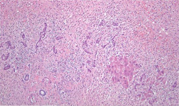
Fig. 1.
Liver explant from patient #1, a 44-year-old female, with massive hepatic necrosis. The only hepatocytes in this field are the small collection to the right of center. The remainder of this field is collapsed parenchyma resulting from confluent lobular necrosis, with a portal tract at the lower left and proliferating bile ductules and neocholangioles to the left of center. (H&E stain, × 100 magnification)

