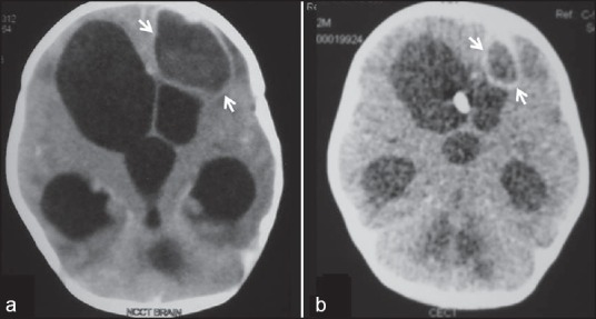
Figure 1.

Contrast enhanced CT scan of the head: Contrast enhanced axial CT scan image of the brain (a) shows a welldefined peripheral enhancing cystic lesion (arrows) in left frontal lobe with surrounding edema consistent with abscess with marked hydrocephalus. Follow-up CT scan after antifungal treatment (b) shows near complete resolution of the abscess
