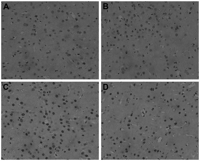
Figure 2.
Apoptosis analysis of cerebral cortex at 24 h (magnification, x200). The (A) control, (B) valproic acid, (C) X-ray and (D) combined groups. Cell apoptosis was analyzed in the cerebral cortex according to the method of immunohistochemistry using protein caspase-3. All micrographs are magnification, x200.

