Figure 1.

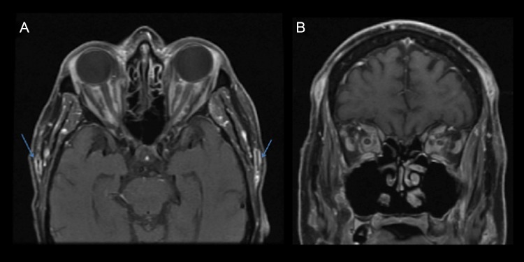
Contrast enhanced MRI orbits. (A) Bilateral enhancement of superficial temporal artery (arrows). (B) Intraconal fat stranding and inflammatory debris.

Contrast enhanced MRI orbits. (A) Bilateral enhancement of superficial temporal artery (arrows). (B) Intraconal fat stranding and inflammatory debris.