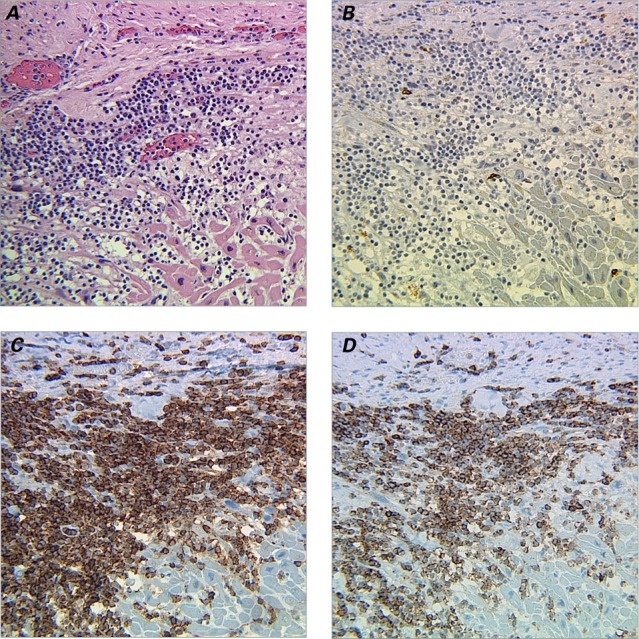
Fig. 2.

Photomicrographs show myocardium infiltrated with T-cell prolymphocytic leukocytes. A) Section shows myocardial fibrosis and compensatory hypertrophy with leukemic cell infiltration (H & E, orig. ×200). Immunohistochemical staining (orig. ×200) reveals B) leukemic cells negative for CD20, but positive for C) CD4 and D) CD8.
